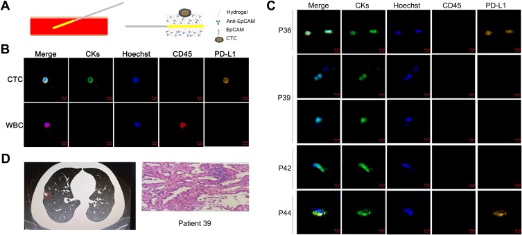
Figure 2.
Collection and determination of CTCs. (A) CellCollector in vivo CTCs detection system. A medical stainless steel wire with a 2-cm-long functional domain coated with EpCAM antibody and a hydrogel stratum. The sampling probe was inserted into cubital vein peripheral blood through a 20G catheter with the functional domain exposed to the peripheral blood and placed in vivo for 30 min to capture tumor cells. (B) Determination of tumor cells by CK7/19/panCK (green channel). Hoechst was used for nuclear counterstaining (blue channel), and white cells were determined by CD45 staining (red channel). PD-L1 expression in CTCs was determined by PD-L1 staining (orange channel). Scale bar: 10 μm. WBC indicates white blood cell. (C) Collection and determination of CTCs in the recruited patients by CellCollector. Two CTCs were collected in patient number 36, 3 CTCs in patient number 39, and 1 CTC in patient numbers 42 and 44. P indicates patient. (D) Imaging and pathological results for patient number 39. A pulmonary nodule was found in the right lung. The patient was diagnosed with invasive adenocarcinoma by pathology.

