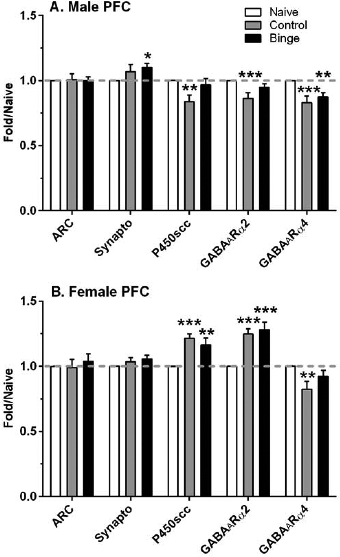
Figure 2. Sex dependent changes in relative density of select proteins in the prefrontal cortex (PFC) after a history of ethanol drinking with repeated intermittent predator odor stress exposure.

Western blot analysis was conducted on dissected PFC tissue at 24 h after the final drinking session (binge = prior binge ethanol intake; control = prior water intake) and compared to values from similarly aged naïve mice (naïve). Sexually divergent effects of treatment were observed on protein levels of synaptophysin (synapto), cytochrome P450 side chain cleavage (P450scc), and GABAA receptor (GABAAR) α2 subunit in male and female PFC, whereas treatment produced a similar suppression in GABAAR α4 subunit protein levels. Values are mean ± SEM for each group: male: n=15–18 (naïve), n=11 (control), n=12 (binge); female: n=13–17 (naïve), n=8 (control), n=10–11 (binge). All levels were initially normalized to β-actin. Fold regulation was then determined by normalizing all values to the mean of the relative expression of the respective naïve group (dashed gray line). *p<0.05, **p<0.01, ***p≤0.001 vs respective naïve group (effect size range = 1.31 – 3.41).
