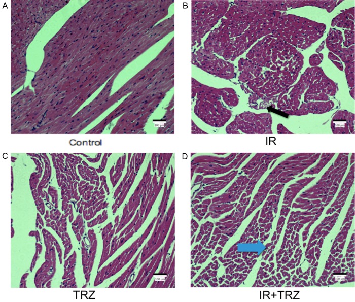
Figure 4.
Histopathology changes in the heart stained with H&E from IR or TRZ treatment after 21 days. A. The normal heart slice was observed in the control group. C. The TRZ group showed relatively normal myocardial structure. B. The black arrow showed the vacuolar degeneration in IR group. D. The blue arrow showed the vacuolar and adipocyte changes in the combined group. The arrangement of myocardial cell was disordered in the combined group, as well as the loose of structure of myocardial cells and the pyknosis of the nucleus.

