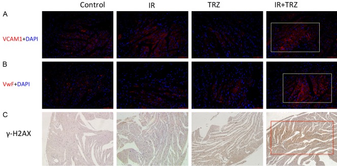
Figure 5.
The microvascular lesions and DNA damage in the heart tissue. (A) VCAM-1 (Red) and (B) vWF (Red) expression on the cardiac slice was detected by immunofluorescence staining. Cell nuclei were labeled by DAPI (blue). (C) The immunohistochemistry staining determines the γ-H2AX expression in the cardiac slice.

