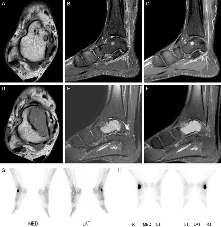
Figure 1.

Radiologic findings. (A-F) Axial T1 weighted (A) and sagittal fat-saturated T2 weighted (B) images show a well-defined 0.8 cm, round mass with intermediate signal intensities eccentrically located in the right talar neck. (C) It shows prominent enhancement on sagittal enhanced fat-saturated T1 weighted image. Axial T1 weighted (D) and sagittal fat-saturated T2 weighted (E) images taken 5 years later show a slightly lobulated expansile 4.4 cm mass in the medial aspect of the talus. (F) The mass shows strong enhancement on sagittal enhanced fat-saturated T1 image. (G, H) Bone scans show focal increased uptake in the right talus initially (G) and an increased extent of hot uptake after 5 years (H), suggesting progression of the bone tumor.
