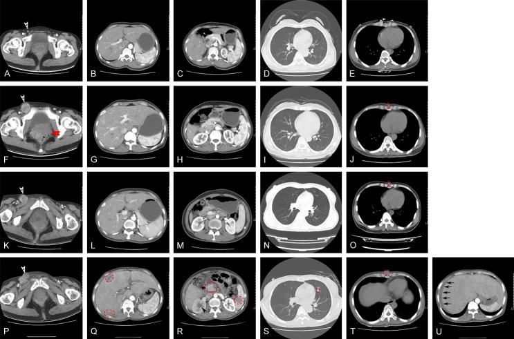
Figure 1.
A: Right inguinal area with multiple lymph nodes (white arrow). B-E: No obvious abnormality was found in a CT scan of liver, stomach, pancreas, spleen, lung, kidney and mediastinum. F: Right inguinal area had multiple enlarged lymph nodes (white arrow), and primary tumor (red arrow). G-I: No obvious abnormality was found in CT scan of liver, stomach, pancreas, spleen, kidney and lung. J: A small nodule appears in front of the xiphoid process (red circle). K: Right inguinal area with multiple enlargement of lymph nodes (white arrow). L-N: No obvious abnormality was found in CT scan of liver, stomach, pancreas, spleen, kidney and lung. O: A small nodule appears in front of the xiphoid process (red circle). P: Right inguinal area multiple enlargement lymph nodes (white arrow). Q: Multiple liver metastases (red circle). R: Pancreas and spleen metastases (red circle). Hydronephrosis of right kidney (black arrow). S: Multiple lung metastases (red circle). T: Soft tissue metastasis in front of the xiphoid process (red circle). U: Ascites (black arrows).

