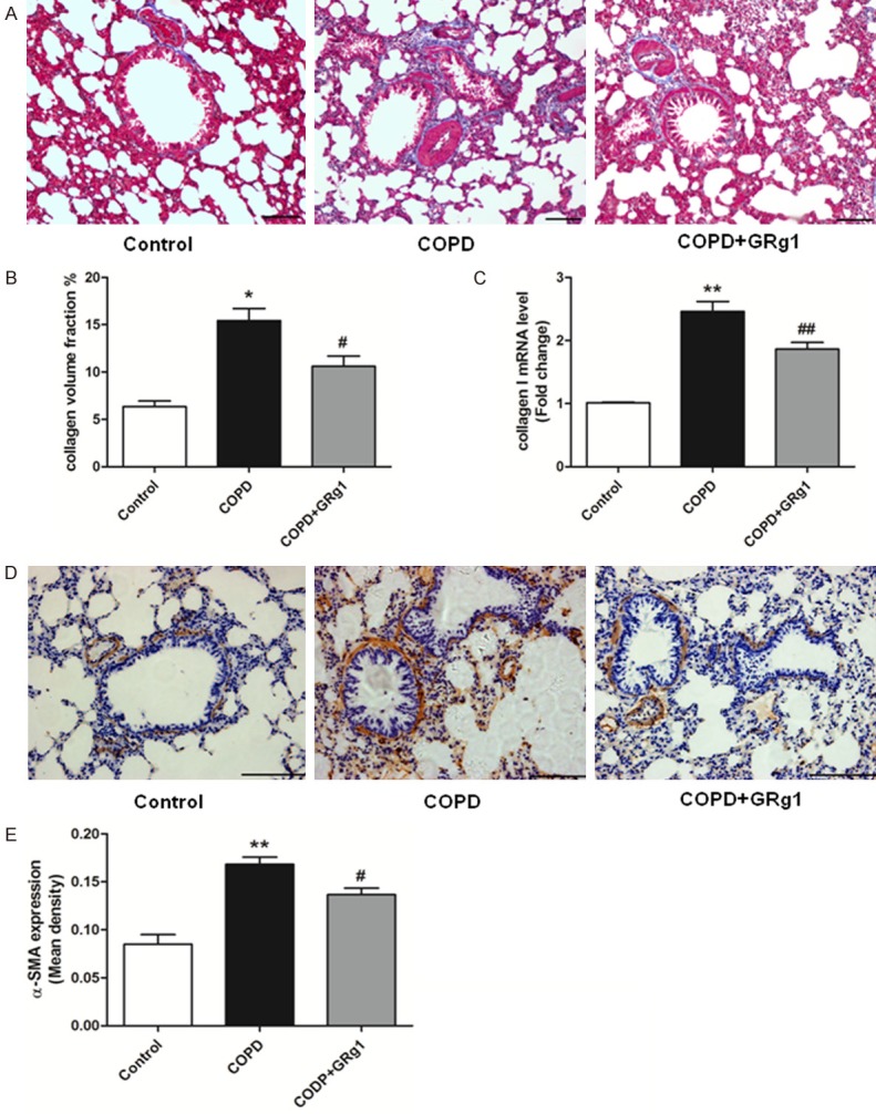
Figure 2.

GRg1 attenuated airway fibrosis in COPD rats. A. At the end of week 12, lung sections were prepared, and Masson Trichrome staining analysis was used to assess the fibrotic changes of lung tissue. N = 6 in each experimental group. Images were taken at ×100 magnification. Scale bar = 100 μm. B. Quantitative collagen assay indicated that GRg1 markedly reduced the ratio of area with collagen accumulation in COPD rats. C. The mRNA level of collagen I was measured by real-time PCR. The results expressed as relative mRNA expression (relative to GAPDH mRNA level). CS exposure promoted mRNA expression of collagen I in lung tissue, and could be attenuated by GRg1. D. Lung sections were prepared, and immunohistochemistry was used to assess the expression of α-SMA. Images were taken at ×200 magnification. Scale bar = 100 μm. E. CS increased the expression of α-SMA in lung tissue, and was abated by GRg1. Data were expressed as mean ± SD. Statistical significance was assessed by one-way ANOVA and Tukey’s post hoc test. *P<0.05 and **P<0.01 vs. control; #P<0.05 and ##P<0.01 vs. COPD.
