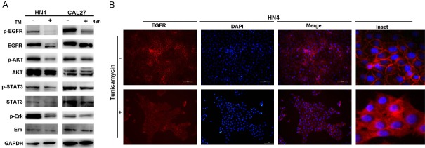
Figure 4.
TM suppresses the EGFR signaling pathway and facilitates the translocation of EGFR. A. Western blot analysis of p-EGFR, EGFR, p-AKT, AKT, p-STAT3, STAT3, p-Erk and Erk protein levels of HN4 and CAL27 cells with or without of TM (2 µg/ml) treatment for 48 hours. B. Image of immunofluorescence staining for EGFR expression in HN4 cells treated with or without TM (2 µg/ml) for 48 hours with nuclei stained with DAPI (blue). Scale bar = 50 μm.

