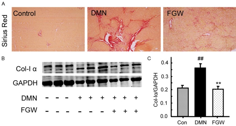
Figure 2.

Evaluation for the collagen deposition in liver tissues of hepatic fibrosis rat. A. Sirius red staining for the hepatic fibrosis. B. Western blot bands for the collagen I expression. C. Statistical analysis for the collagen I expression. ##P < 0.01 vs. Control group. **P < 0.01 vs. DMN group.
