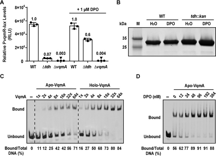
Figure 1.
VqmA is active in the absence of DPO. A, PvqmR-lux reporter activity for the indicated V. cholerae strains after 6 h of growth in the absence or presence of 1 μm DPO. The cells were at HCD at this time point. Data are represented as mean ± S.D. (error bars) with n = 3 biological replicates. B, SDS-polyacrylamide gel of purified His-VqmA protein produced in WT or tdh::kan E. coli without or with 100 μm DPO added during expression. M, molecular weight marker. C, EMSA showing VqmA binding to PvqmR promoter DNA. 540 pm biotinylated PvqmR DNA and either no protein (lane 1, −), apo-VqmA (lanes 2–8), or holo-VqmA (lanes 9–15). Relative protein concentrations are indicated: 1× = 2 nm and 64× = 130 nm. D, EMSA showing binding of 50 nm apo-VqmA (lanes 2–8) to PvqmR DNA in the absence and presence of the indicated amounts of DPO. Lane 1, as in C. In C and D, quantitation of the percentage of PvqmR DNA bound appears below each lane. RLU, relative light units.

