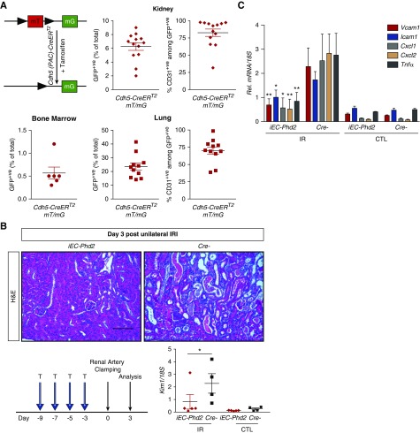
Figure 6.
Inducible endothelial Phd2 inactivation is sufficient to attenuate renal IRI and postischemic inflammation. (A) Cdh5(PAC)-CreERT2 mice were crossed to ROSA26-ACTB- tdTomato,-EGFP reporter (mT/mG) mice such that injection of tamoxifen in adult life results in GFP expression that is highly selective for ECs. The degree of EC recombination was assessed by FACS analysis in single-cell suspensions prepared by BM, kidney, and lung. Shown is the percentage of GFP+ cells in BM (n=6), kidney (n=13), and lung (n=11). Right panels show the percentages of CD31+ cells contained with the GFP+ cells in kidneys (top) and lungs (bottom). (B) Experimental scheme illustrating the timing of tamoxifen administration, renal artery clamping, and analysis. Representative images of hematoxylin and eosin (H&E)–stained sections from postischemic kidneys and Kim1 mRNA levels in IR and CTL kidneys from iEC-Phd2 mice or their Cre- controls at day 3 after unilateral renal IRI (n=5–4, P=0.046; one-way ANOVA with Sidak correction for multiple comparisons). (C) Quantitative RT-PCR analysis for Vcam1, Icam1, Cxcl1, Cxcl2, and Tnfa in IR and CTL kidneys of mice with the indicated genotypes at day 3 after unilateral renal IRI (n=5–4, Vcam1 P=0.008, Icam1 P=0.05, Cxcl1 P=0.023, Cxcl2 P=0.002, Tnfa P=0.009; one-way ANOVA with Sidak correction for multiple comparisons). Error bars represent SEM. *P<0.05, **P<0.01. Scale bars, 100 μm. CTL, contralateral kidney; IR, kidney subjected to unilateral IRI; Rel., relative; T, tamoxifen.

