Figure 7.
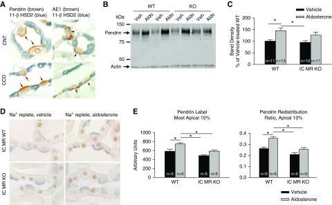
IC MR gene ablation blunts, but does not eliminate, aldosterone’s effect on apical pendrin label and subcellular distribution. (A) 11β-HSD2 immunolabel (blue) in AE1- and pendrin-positive ICs (brown) and in AE1-/pendrin-negative principal cells or CNT cells from the CNT and CCD. As shown, no 11β-HSD2 label was observed in ICs from either segment. (B) Pendrin total protein abundance in a representative immunoblot of kidney lysates from aldosterone- and vehicle-treated wild-type and IC MR null mice. (C) Pendrin band density in kidney lysates from each group shown in (B). (D) Pendrin (blue) and MR (brown) immunolabel in sections of cortical labyrinth of aldosterone- and vehicle-treated wild-type and IC MR null mice. (E) The effect of aldosterone on pendrin’s abundance in the most apical 10% of ICs as well as pendrin label in the most apical 10% of the cell relative to label across the entire cell (pendrin redistribution ratio) in the CNT of wild-type and IC MR null mice. In immunoblots, groups were compared by ANOVA with a Tukey post-test. In immunohistochemistry studies, groups were compared by ANOVA with a Hochberg post-test. *P<0.05.

