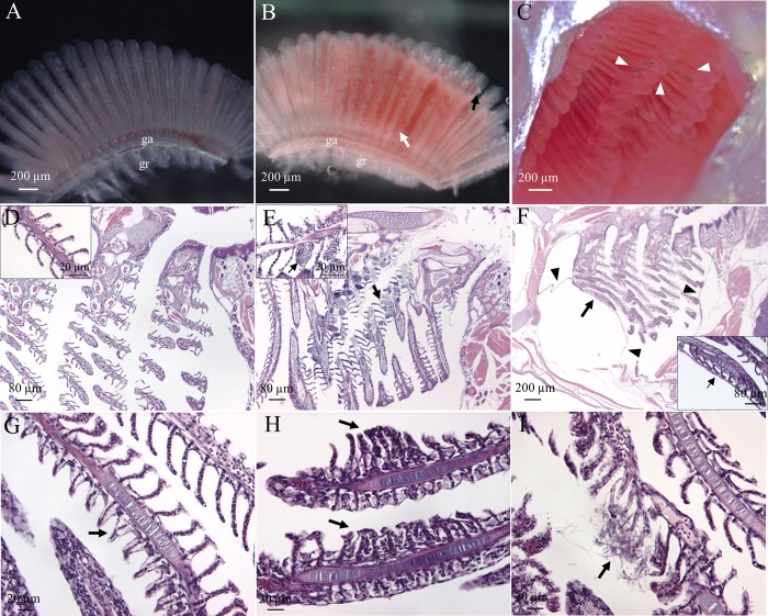
Fig 4. Gill alterations following MF exposure.
Light micrographs of gills from control (A) and PES-exposed (B) medaka after 2 days of exposure; white arrow indicates aneurysms, black arrow indicates normal lamellar outgrowths, arrowheads indicate PES MFs in the branchial cavity (C). H&E stained histological sections of gills from the adult medaka exposed to 0 (D), PES (E-G, I), or PP (H) MFs for 21 days. (E) Black arrows under low and high magnifications of the filaments indicate aneurysms. (F) Arrowheads indicate swelling between deep layers of the operculum associated with the wall of the branchial chamber and arrows show pushing of inner opercular epithelium against gill primary lamellae, visible in more detail in high magnification inset. (G) Arrow indicates epithelial lifting in the secondary lamellae. (H) Arrows indicate fusion of secondary lamellae. (I) Arrow indicates epithelial alterations of the secondary lamellae. ga, gill arch; gr, gill raker.

