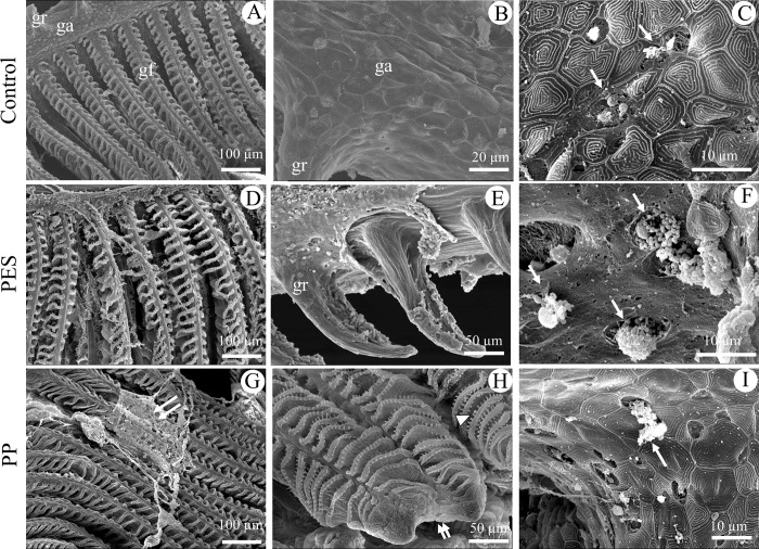
Fig 5. SEM of gills after 21 days of exposure.
0 (Control; A-C), PES (D-F), or PP (G-I) MFs. (A, D, G) gill filaments, only a portion of one gill raker may be observed in control figure (A). (B) Gill arch. (E) Gill raker. (C, F, I) Magnification of gill arch showing mucous cells indicated by white arrows. (G) Double white arrow indicates mucous secretion as a sheet; (H) Magnification of the filament tip with arrowhead to outgrowth and showing fusion of distal tips of adjacent primary lamellae (double white arrow). ga, gill arch; gf, gill filament; gr, gill raker.

