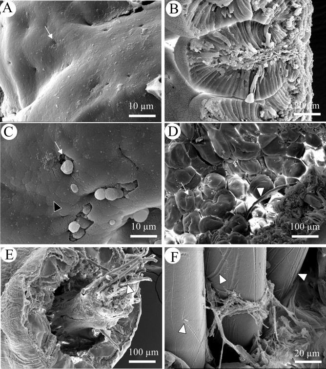
Fig 6. SEM of cross sections of gut.
(A) Surface epithelium of foregut from a control fish with white arrows marking pores for mucous secretion; (B) Transverse section of hindgut from control fish; (C) Surface epithelium of foregut from PES-exposed fish, black arrowhead indicates apical tips of enterocyte, white arrow indicates mucus secretion; (D) Low magnification of foregut from PES-exposed fish with fiber entangled in folds (white arrowhead); (E) Low magnification of hindgut from PES-exposed fish with fibers (white arrowhead) encased in digesta; (F) High magnification of PP in hindgut showing elongated grooves on their surfaces.

