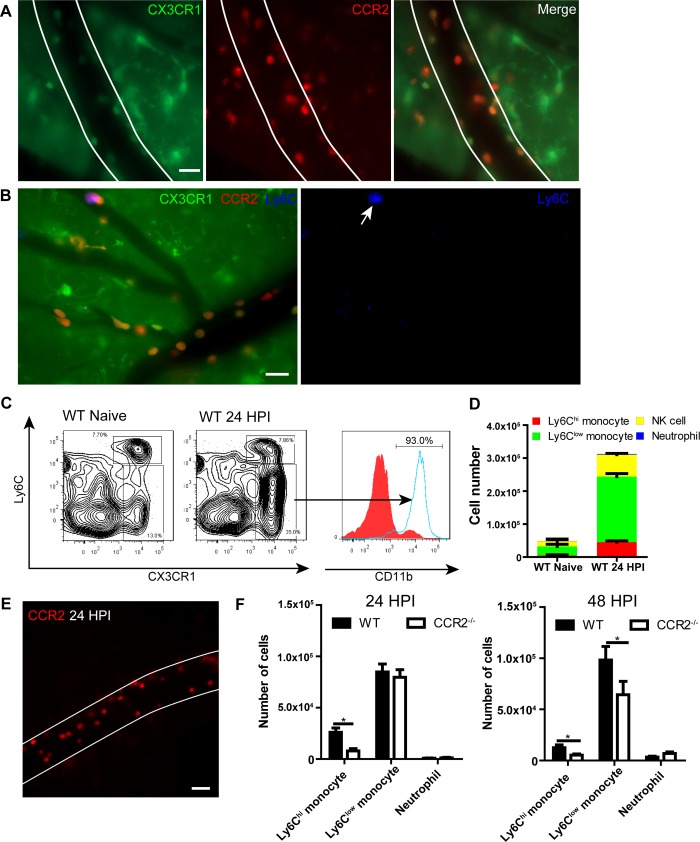
Fig 2. Recruited monocytes are mainly Ly6Clow populations.
(A) IVM images showing the expression of CX3CR1 and CCR2 on recruited monocytes in brain postcapillary venules of CX3CR1gfp/+CCR2rfp/+ mice 24 h after infection with 20x106 C. neoformans (Green: CX3CR1, Red: CCR2). (B) Expression of Ly6C on recruited monocytes (arrow) in brain postcapillary venules of CX3CR1gfp/+CCR2rfp/+ mice 24 h after infection with 20x106 C. neoformans. For labeling of Ly6C, mice were i.v. injected with 2 μg AF647-anti-Ly6C mAb through the tail vein 5 min before imaging. (C) Representative flow cytometry plots of CX3CR1+Ly6Chi and CX3CR1+Ly6Clow monocyte subsets in the brain of naïve and infected C57BL/6 mice. Gated on CD45+ cells. The expression of CD11b on CX3CR1- (filled) and CX3CR1+ (solid) populations was shown on the right. C57BL/6 mice were i.v. infected with 20x106 C. neoformans and 24 h later brain leukocytes were isolated and analyzed by flow cytometry. (D) Flow cytometry analysis of the numbers of different subsets of cells recruited to the brain of C57BL/6 mice (n = 4 per group) 24 h after infection. Ly6Chi monocytes were defined as CD45+Ly6G-NK1.1-CD11b+CX3CR1+Ly6Chi, Ly6Clow monocytes as CD45+Ly6G-NK1.1-CD11b+CX3CR1+Ly6Clow, NK cells as CD45+NK1.1+, neutrophils as CD45+CD11b+Ly6G+. (E) A representative IVM image showing the recruitment of RFP+ monocytes in brain postcapillary venules of CCR2rfp/rfp mice (CCR2 deficient) 24 h after i.v. infection with 20x106 C. neoformans. See also S4 Video. (F) Flow cytometry analysis of the numbers of Ly6Chi and Ly6Clow monocytes and neutrophils recruited to the brain of WT and CCR2-/- mice (n = 4 per group) 24 and 48 h post i.v. infection with 20x106 C. neoformans. Scale bars: 20 μm. Data are expressed as mean ± SEM and representative of 3 independent experiments. * p<0.05.

