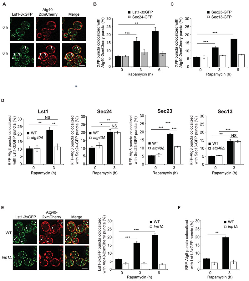
Figure 2. Lst1 and Sec23 colocalize with Atg40 and Atg8 in rapamycin-treated cells.
(A) Representative images of cells treated for 0 or 6 h with rapamycin. Arrowheads indicate Lst1-3xGFP that colocalize with Atg40-2xmCherry puncta. (B) Bar graph shows the percent of Lst1-3xGFP or Sec24-GFP colocalizing with Atg40-2xmCherry puncta. (C) Bar graph shows the percent of Sec23-GFP or Sec13-GFP colocalizing with Atg40-2xmCherry puncta. (D) Cells were treated for 0 or 3 h with rapamycin, and RFP-Atg8 that colocalizes with GFP tagged COPII coat subunits was quantitated. (E) Left, arrowheads indicate Lst1-3xGFP that colocalizes with Atg40-2xmCherry puncta 6 h after rapamycin treatment. Quantitation is on the right. (F) Quantitation of RFP-Atg8 that colocalize with Lst1-3xGFP in WT and lnp1Δ cells. Scale bars in (A), (E), 5μm. Error bars in (B-F) represent S.E.M., N=3-6; NS, not significant P ≥ 0.05, *P < 0.05, **P < 0.01, ***P < 0.001, Student’s unpaired t-test.

