. 2020 Feb 13;9:e53865. doi: 10.7554/eLife.53865

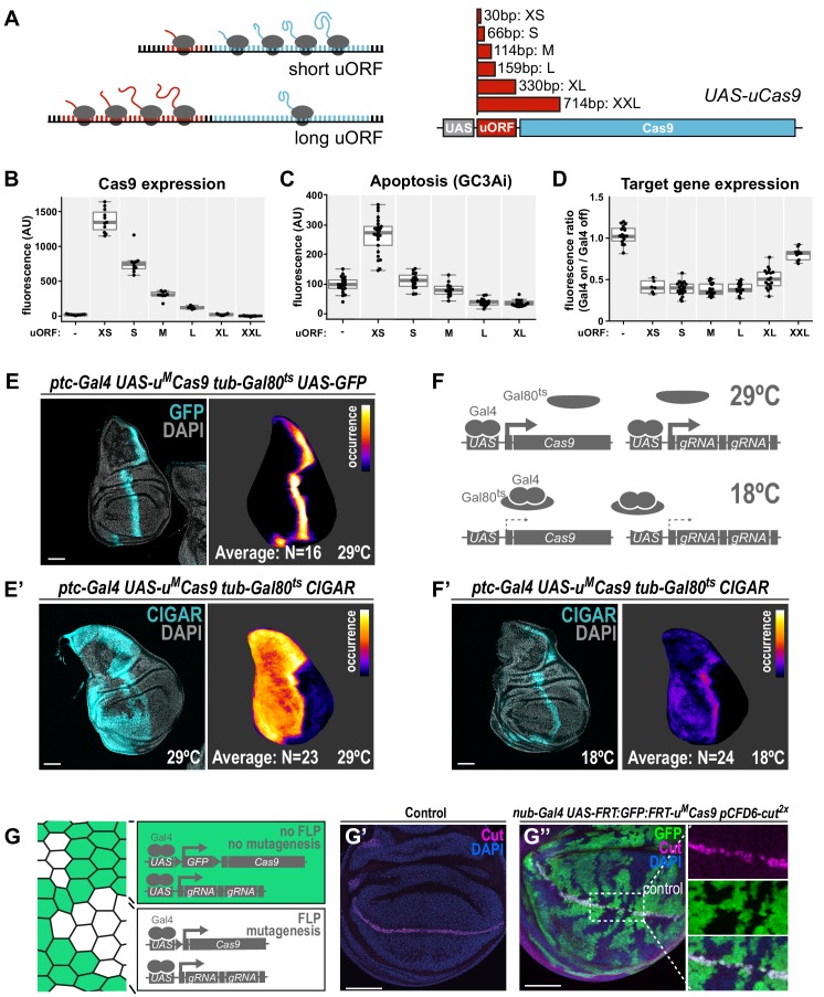
Figure 2. A transgenic series for tunable Cas9 expression to balance activity and toxicity.

(A) Principle of the UAS-uCas9 series. Translation of the downstream ORF is inversely correlated with length of the upstream ORF in bicistronic mRNAs. The UAS-uCas9 series consists of transgenes that harbor uORFs of different length to modulate expression of Cas9. (B - D) Systematic characterization of Cas9 expression, toxicity and mutagenesis efficiency of the UAS-uCas9 series. Transgenes of the UAS-uCas9 series were recombined with nub-Gal4 and crossed to the apoptosis sensor UAS-GC3Ai (B, C) or pCFD6-evi2x (D). Graphs show data as individual dots, and boxplots as a data summary, with the line representing the median and the box the interquartile range. (B) Quantification of anti-Cas9 staining intensity in wing discs of the indicated genotype. Cas9 levels gradually reduce as the size of the uORF increases. N ≥ 6 wing discs. (C) Elevated levels of apoptosis were only observed with UAS-uXSCas9. The longest uORF (uXXL) encodes EGFP, preventing visualization of dying cells with GC3Ai. Quantification of fluorescent intensity of the GC3Ai reporter in the wing pouch. N ≥ 14 wing discs. (D) All transgenes of the UAS-uCas9 series mediate evi mutagenesis, with transgenes containing the four shortest uORFs (XS-L) leading to comparable gene editing that removes Evi from nearly all cells in the Gal4 expression domain. Quantification of staining intensity for Evi protein in the wing pouch (Gal4 on), relative to Evi staining in the hinge region (Gal4 off). N ≥ 6 wing discs. (E, E’) CRISPR mutagenesis patterns reflect Gal4 expression history. (E) Fluorescence of GFP, which turns over, reflects most recent Gal4 expression pattern. (E’) CRISPR mutagenesis, visualized by activation of the CIGAR reporter, is permanent and reveals the Gal4 expression history. Images of a representative wing disc are shown to the left of each panel and average intensity projection of several discs registered to a common template are shown on the right (see Materials and methods). Areas that are CIGAR positive in many discs appear bright, while areas devoid of signal in most discs appear dark. (F, F’) Incomplete repression of CRISPR mutagenesis by temperature-sensitive Gal80. (F) Principle of the Gal80ts system. At 18°C Gal80 binds and inhibits Gal4. (F’) Mutagenesis is still observed at 18°C in 11/24 discs and observed preferentially in the Gal4 expression domain, indicating incomplete Gal4 suppression by Gal80ts. (G - G’’) Control of CRISPR mutagenesis by a flip-out cassette. (G) In the absence of FLP recombinase a FRT-flanked GFP flip-out cassette (FRT sites represented by triangles) separates Cas9 from the promoter, resulting in cells that express GFP, but no Cas9. In the presence of FLP, the GFP cassette is excised and Cas9 is expressed. (G’) Staining for the transcription factor Cut reveals a continuous stripe of cells expressing ct along the dorsal-ventral boundary in wildtype tissue. (G’’) A pulse of FLP expression was used to excise the GFP flip-out cassette in a subset of cells (marked by the absence of GFP). Cut expression (magenta) is exclusively lost in GFP negative cells. Scale bar = 50 µm.

Figure 2.

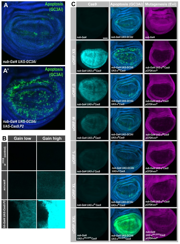
Figure 2—figure supplement 1. High levels of Cas9 expression from UAS-cas9.

Figure 2—figure supplement 1.

P2 are cytotoxic. (A) Expression of Cas9 in nub-Gal4 UAS-Cas9.P2 causes excessive cell death. Apoptotic cells were visualized by co-expression of GC3Ai, which is fluorescent in cells with activated caspase 3. Expression of GC3Ai in the wing pouch highlights a few cells undergoing apoptosis. Additional expression of UAS-cas9.P2 causes a dramatic increase in the cells undergoing programmed cell death, highlighting cytotoxicity caused by high levels of Cas9. (B) Comparison of Cas9 expression levels between act-cas9 and hh-Gal4 UAS-cas9.P2 wing discs. Cas9 was detected by antibody staining and imaged in the same session with identical setting at either low (top panel) or high (bottom panel) detector gain. The difference in Cas9 levels is such, that Cas9 expressed from act-cas9 is undetectable with low detector gain and Cas9 staining in hh-Gal4 UAS-cas9.P2 wing discs is oversaturated with high detector gain, where Cas9 from act-cas9 is visible. The act-cas9 transgene is known to mediate highly efficient mutagenesis in Drosophila (Port et al., 2014; Port et al., 2015 ). (C) Systematic characterization of Cas9 expression, toxicity and mutagenesis of the UAS-uCas9 series. Transgenes of the UAS-uCas9 series were recombined with nub-Gal4 and wing imaginal discs were stained for Cas9 protein. Cas9 levels gradually reduce as the size of the uORF increases (left panel). nub-Gal4 UAS-uCas9 flies were crossed to UAS-GC3Ai to visualize cells undergoing apoptosis. Elevated levels of apoptosis were only observed with UAS-uXSCas9. The longest uORF (uXXL) encodes EGFP, preventing visualization of dying cells with GC3Ai (middle panel). nub-Gal4 UAS-uCas9 flies were crossed to pCFD6-evi2x and mutagenesis of evi was indirectly observed by loss of Evi staining. All transgenes of the UAS-uCas9 series mediate evi mutagenesis, with transgenes containing the four shortest uORFs (XS-L) leading to comparable gene editing that removes Evi from nearly all cells in the Gal4 expression domain (right panel).
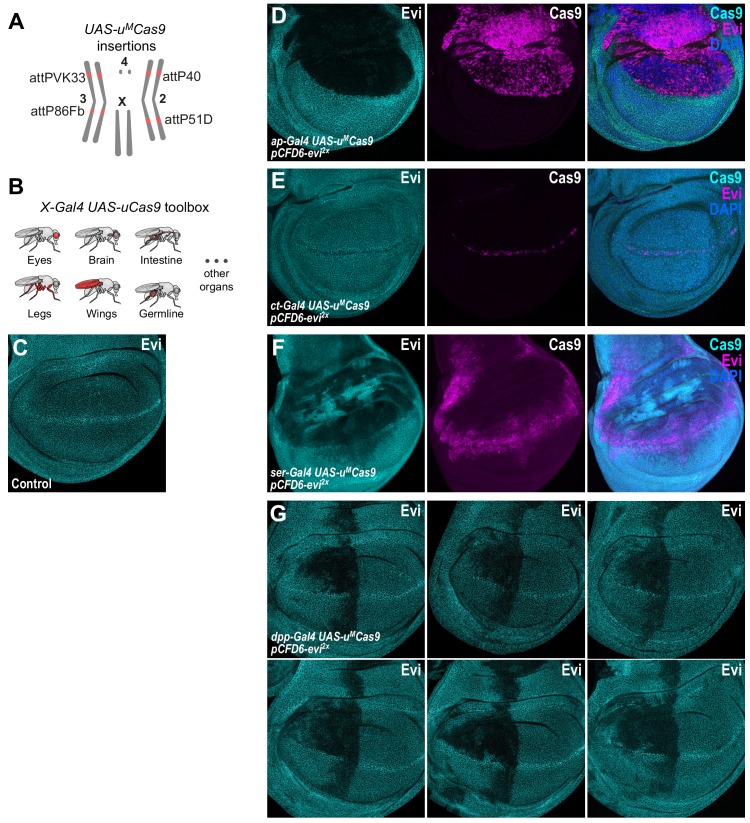
Figure 2—figure supplement 2. CRISPR mutagenesis patterns reflect expression patterns of Gal4 lines throughout development.

Figure 2—figure supplement 2.

(A) Chromosomal locations of UAS-uMCas9 transgenes currently available. (B) A growing collection of Gal4 UAS-uCas9 stocks will allow tissue-specific mutagenesis in Drosophila. (C) Evi is expressed in all cells of wildtype third-instar wing imaginal discs and accumulates in cells at the dorsal-ventral boundary. (D) Mutagenesis of evi in ap-Gal4 UAS-uMCas9 pCFD6-evi2x wing discs results in loss of Evi protein exclusively in the dorsal compartment, which expresses Cas9 protein. (E) CRISPR mutagenesis of Evi with ct-Gal4 results in loss of Evi staining only in cells along the dorsal-ventral boundary, where also Cas9 protein is expressed. (F) CRISPR mutagenesis with ser-Gal4 results in loss of Evi staining in the pouch, hinge and notum region of the wing disc and includes large areas where no Cas9 protein is detectable in third instar wing discs. (G) Mutagenesis of evi with dpp-Gal4. Dpp is known to be expressed in a stripe along the anterior-posterior boundary in third instar wing imaginal discs. In addition to this domain, mutagenesis is consistently found in the anterior-dorsal region of the pouch. Six representative discs for the same genotype are shown.