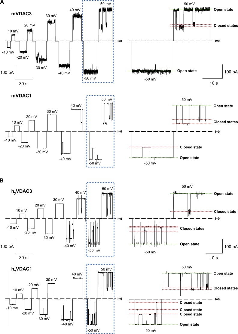
Figure S3.
Recombinant mouse VDAC3 and VDAC1 isolated from E. coli inclusion bodies (mVDAC3 and mVDAC1) and human VDAC3 and VDAC1 isolated from yeast mitochondria (hyVDAC3 and hyVDAC1) form typical VDAC channels. (A and B) Representative single-channel current traces obtained with reconstituted mVDAC3 and mVDAC1 expressed in E. coli and refolded from inclusion bodies (A), and hyVDAC3 and hyVDAC1 expressed in yeast and isolated from mitochondria (B), at different applied voltages. Framed traces are displayed in fine time scale on the right. Dashed line indicates zero current level and dotted lines indicate VDAC’s open and closed states. Current records were digitally filtered at 100 Hz using a low-pass Bessel (8-pole) filter. Membrane-bathing solutions consisted of 1 M KCl buffered with 5 mM HEPES at pH 7.4. Planar membranes were formed from soybean PLE with 5% cholesterol (wt/wt).

