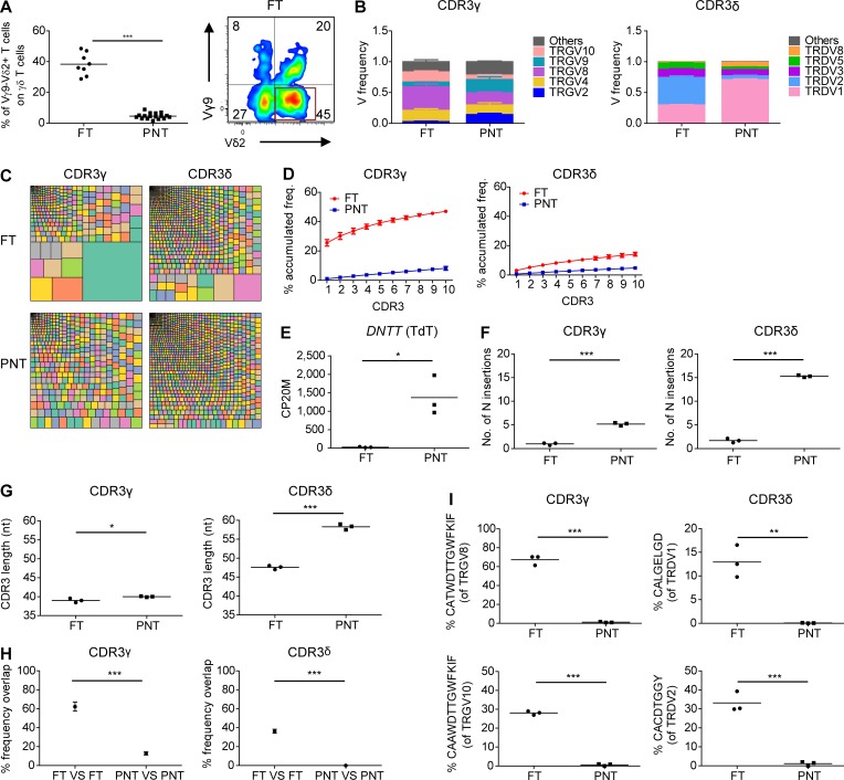
Figure 2.
Human fetal γδ thymocytes express invariant germline-encoded CDR3γ and CDR3δ repertoires. (A) Flow cytometry analysis of the expression of Vγ9−Vδ2+ γδ T cell subset in FT and PNT γδ thymocytes (left); representative flow cytometry plot of fetal γδ thymocytes (right). (B) HTS analysis of the V gene segment usage TRGV (left) and TRDV (right) in FT and PNT γδ thymocytes. TRG “Others” groups: TRGV1-TRGV3, TRGV5, TRGV5P, TRGV7, and TRGV11 variable chains; TRD “Others” groups: TRDV4, TRDV6, and TRDV7 variable chains. (C) Tree map representations of the CDR3γ and CDR3δ repertoires of FT and PNT γδ thymocytes (colors used are random; no correspondence between graphs). Representative of three independent experiments. (D) Accumulated frequencies (freq.) of the 10 most abundant clonotypes in CDR3γ and CDR3δ repertoires of γδ thymocytes in FT and PNT. (E) Analysis of the “counts per 20 million” (CP20M; RNAseq data) of the DNTT gene of γδ thymocytes in FT and PNT. (F and G) Mean number of N insertions (F) and CDR3 length (G; number of nucleotides) in CDR3γ and CDR3δ repertoires of γδ thymocytes in FT and PNT. (H) Overlap analysis showing the percentage of shared CDR3γ and CDR3δ clonotypes among three FT γδ thymocytes and among three PNT γδ thymocytes. (I) Percentage of particular clonotype amino acid sequences in CDR3γ and CDR3δ repertoires in FT and PNT γδ thymocytes. Each symbol (A, E–G, and I) represents an individual donor. Graphs show the means ± SEM (B, D, and H) or the mean (A, E–G, and I). Data were analyzed by Student’s t test; *, P < 0.05; **, P < 0.01; ***, P < 0.001. n = 8 (FT) and 17 (PNT; A) or n = 3 (FT and PNT; B–I).

