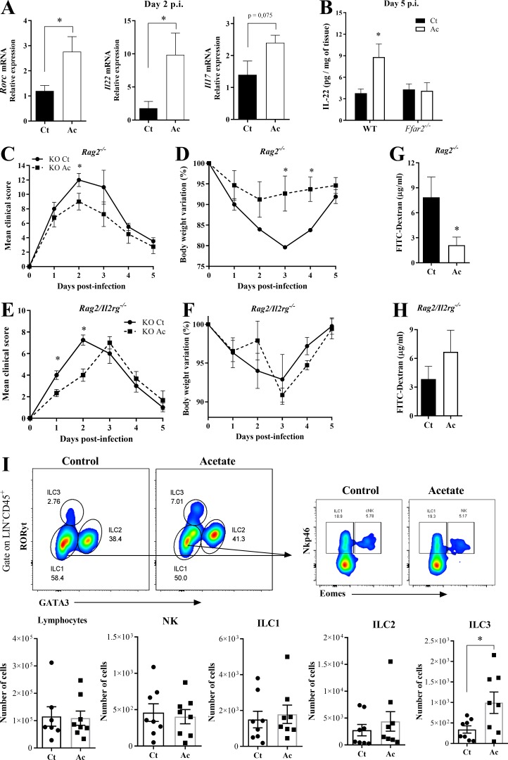
Figure 5.
The protective effect of acetate against CDI depends on ILC3s. (A) Rorc, Il22, and Il17 mRNA expression in the colon of WT mice on day 2 p.i. Mice were treated (Ac) or not (Ct) with acetate and infected with C. difficile (n = 5). (B) IL-22 content in the colon of WT and Ffar2−/− mice on day 5 p.i. Mice were treated or not with acetate and infected with C. difficile (n = 5–6). (C–F) Clinical score and weight changes of Rag2−/− mice (C and D) and Rag2/Il2rg−/− mice (E and F) that were treated or not with acetate and infected with C. difficile (n = 4). (G and H) Intestinal permeability of Rag2−/− (G) and Rag2/Il2rg−/− (H) mice treated or not with acetate on day 2 p.i. (n = 4). (I) Percentages of total lymphocytes, natural killer (NK) cells, ILC1s, ILC2s, and ILC3s in the colon of mice 5 d p.i. (n = 7–8). A representative flow cytometry plot depicting the strategy for identifying NK-ILC subsets is presented at the top. Results are pooled results from two independent experiments with three to six mice in each group (A, B, and I). Results are presented as mean ± SEM. *, P < 0.05.

