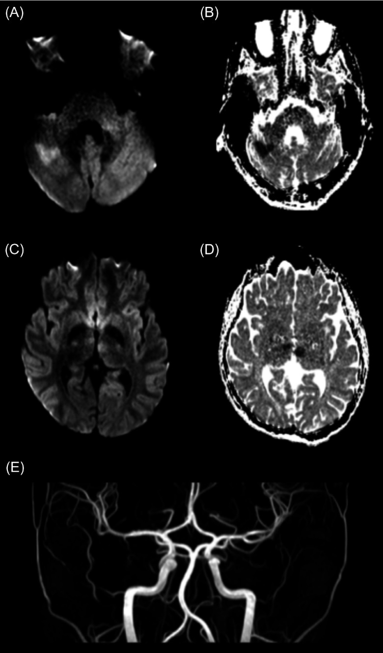
Figure 1:
Diffusion-weighted imaging (DWI) and ADC images acquired 1 h and 10 min after presenting to hospital, approximately 1 hr and 35 min after onset of symptoms, demonstrating areas of schema in the right cerebellar hemisphere and both the right and left thalami. (A) DWI at the level of the cerebellum demonstrating diffusion restriction in the right cerebellar hemisphere. (B) Corresponding ADC demonstrating lower ADC values in corresponding areas to the diffusion restriction on DWI. (C) DWI at the level of the thalami demonstrating diffusion restriction in the right and left thalamus. (D) Corresponding ADC demonstrating lower ADC values in the corresponding areas to the diffusion restriction DWI. (E) MRA demonstrating patency of all arteries.

