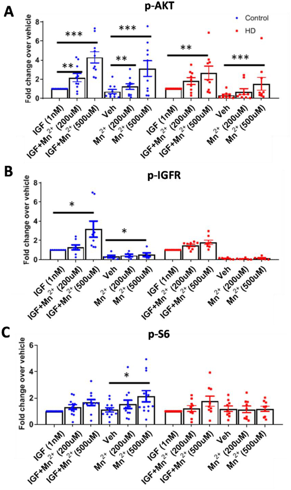
Figure 7: Mn2+-induced p-AKT, IGFR is reduced in HD hiPSC-derived neuroprogenitors.
A-C) Western blot quantification of hiPSC-derived neuroprogenitors from three control patients and three HD patients (CAG repeat 58, 66, 70) for p-AKT (A), p-IGFR (B), and p-S6 (C), following treatment 3hr treatment with Mn2+ (200/500μM) or Mn2++IGF (1nM) in growth factor/insulin free media, after 3hr serum deprivation. N=8–12 for control from three separate patients, N=7–9 for HD including three separate patients; Error bars= SEM. All data normalized to respective control or HD treated IGF-1 alone. Representative blot in Supplemental Fig 5A,B. One-way ANOVA stats listed in Supp Fig 4D. *= significance from vehicle by Dunnet’s multiple comparison test. *P<.05, **P<.01, ***P<.001.

