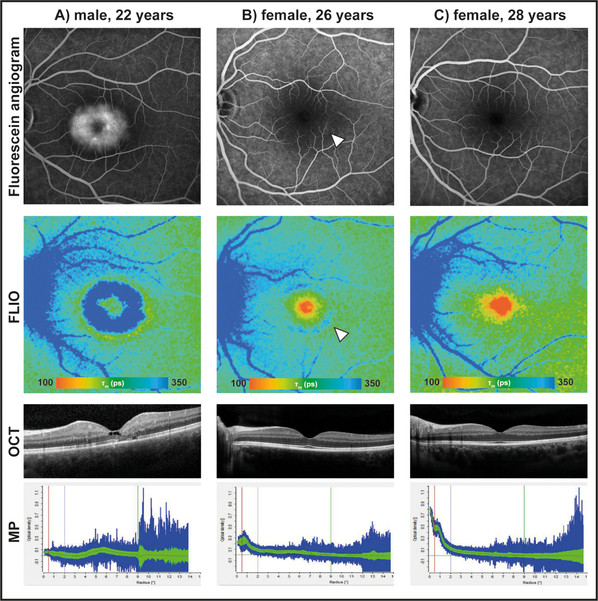
Figure 2: Fluorescein angiograms, fluorescence lifetime (FLIO) images, OCT images, and macular pigment (MP) measurements of three siblings.
A) is affected with MacTel. B) shows FLIO+ changes indicative of MacTel even though her fundus examination was normal, and her other images were graded as just “suspicious” by the reading center due to low macular pigment and faint late-phase inferotemporal fluorescein leakage (arrow). C) shows healthy features of their unaffected sibling.

