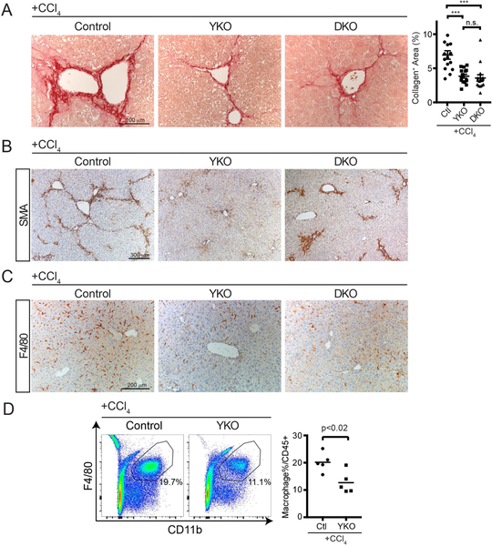
FIGURE 5. Loss of Hepatocyte YAP or YAP/TAZ attenuates Liver Fibrosis after CCl4 Injury.
A. Representative Picro-Sirius Red staining of mice from the indicated genotypes after chronic CCl4 treatment. Dot plot to the right indicates quantification of collagen staining per unit area.
B. Representative αSMA staining of mice from the indicated genotypes after chronic CCl4 treatment.
C. Representative F4/80 staining of mice from the indicated genotypes after chronic CCl4 treatment.
D. Representative flow cytometry of liver macrophages one day after chronic CCl4 treatment. Percentages in the graph are of the gated population. Dot plot to the right of all performed experiments (n=5, each). Horizontal line represents the mean, each dot represents an experiment. Bars above each graph indicate standard error of the mean. **p<0.01, ***p<0.001.

