Fig. 2.
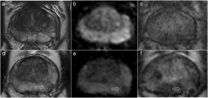
52-year-old patient on active surveillance for Gleason 3 + 3 (1 mm) in the right midgland peripheral zone and a presenting PSA of 6.02 ng/ml (PSA density, 0.12). The first 3-T MRI scan (a–c) did not show any focal lesion but only some patchy diffuse low T2-signal (a) and mild enhancement in the peripheral zone on the right (c) but no restricted diffusion on the ADC map (b). The scan after two years (d–f) revealed a new focal area (arrows) of low T2-signal (d), restricted diffusion on the ADC map (e) and mild enhancement (f) in the left peripheral zone, with a PSA of 8.89 ng/ml (PSA density, 0.18). The PRECISE score was 4 for both radiologists, and targeted biopsy of the area revealed Gleason 3 + 3 (3 mm)

