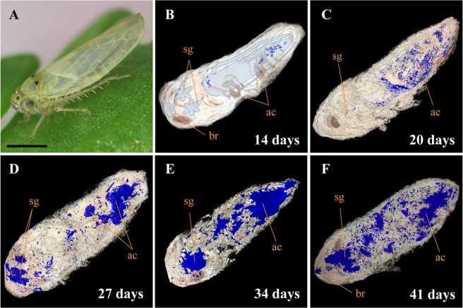
Figure 1.
Distribution of onion yellows (OY) phytoplasmas in leafhopper vector Macrosteles striifrons by three-dimensional (3D) imaging technology. (A) Macrosteles striifrons vector. (B–F) 3D visualization of OY phytoplasmas (blue signals) in its vector, collected at 14 (B), 20 (C), 27 (D), 34 (E), and 41 (F) days after acquisition start (daas) on diseased plants, based on immunohistochemical staining using anti-Amp primary antibody and alkaline-phosphatase-conjugated secondary antibody. The alimentary canal, salivary glands, and brain of the insect are outlined in panel B. ac, alimentary canal; sg, salivary gland; br, brain. Bar, 1 mm.

