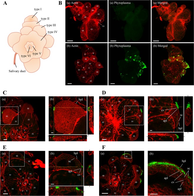
Figure 4.
Distribution of OY phytoplasmas in salivary glands of M. striifrons. (A) Diagrammatic outline of the principal salivary gland of M. striifrons. (B–F) Whole-mount immunofluorescence staining of salivary glands using Amp-Alexa Fluor 488 (green) and the actin dye phalloidin-Alexa Fluor 546 (red). (B) Salivary glands of a non-infected leafhopper (a) and an OY-infected leafhopper that was consistently maintained on a diseased plant for more than 1 month (b). (C–F) Salivary glands of OY-infected leafhoppers at 14 (C), 21 (D), and 28 (E,F) daas. Images in panels C–F are magnified images of boxed areas in image a in the same panel. Image b in each of the panels (C–E) was obtained as described in Fig. 3. All images are representative of more than three experiments. I–VI refers to type I–VI cells; bpl, basal plasmalemma; sc, salivary cytoplasm; apl, apical plasmalemma; cv, cavity. Bar, 75 µm (image a in B), 50 µm (image b in B, image a in F), 25 µm (image a in C–E), 10 µm (image b in C–F).

