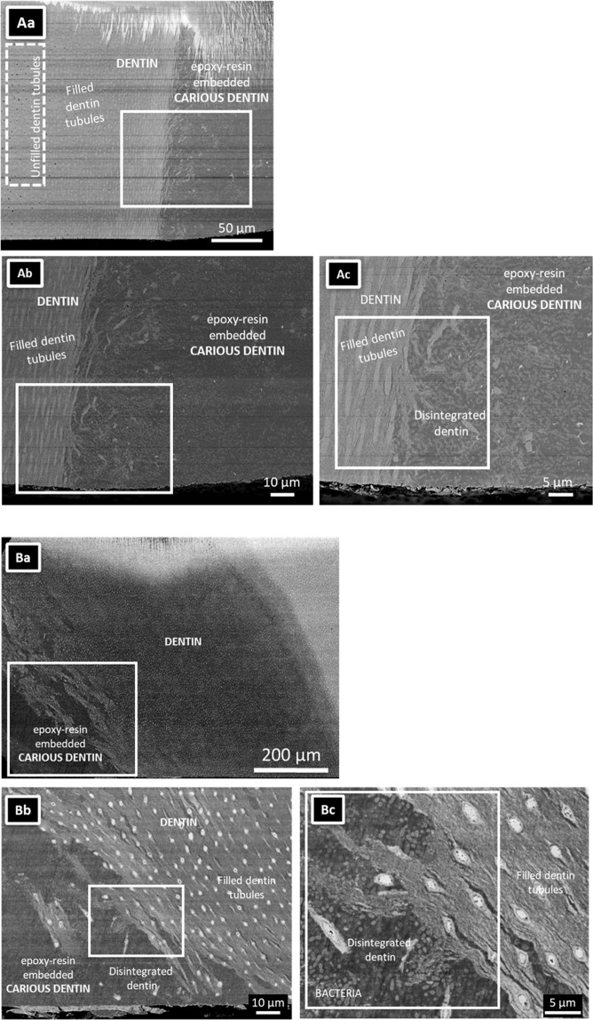
Figure 1.
SEM photomicrographs of a single section through two arrested caries lesions originating from two different carious teeth. (Aa,Ba) Overview SEM photomicrographs of the dentinal caries lesions, illustrating that the caries lesion extended into deep dentin exhibiting many parallel running dentin tubules. The specimens were embedded in epoxy resin. The dotted line demarcates the transition of the caries lesion to the adjacent caries-affected dentin. Note that the specimen top part was damaged by the argon-ion beam of the cross-section polisher (JEOL). (Ab,c) High-magnification SEM photomicrographs revealed open dentin tubules relatively far away from the dentinal caries lesion. Clearly filled dentin tubules were observed near the interface (up to about 100 μm remote from the interface). (Ac) High-magnification photomicrographs confirming that the dentin tubules near the interface with the caries lesion were filled with highly dense material. Collapsed dentin with bacteria was observed near the dentin-caries interface. (Ba) A deep caries region was observed on the lower left of the image. (Bb) High-magnification of (Ba). Collapsed collagen was observed at the interface of the caries lesion with dentin. Filled dentin tubules were observed. (Bc) High-magnification SEM imaging revealing the presence of a considerable number of bacteria at the caries lesion.

