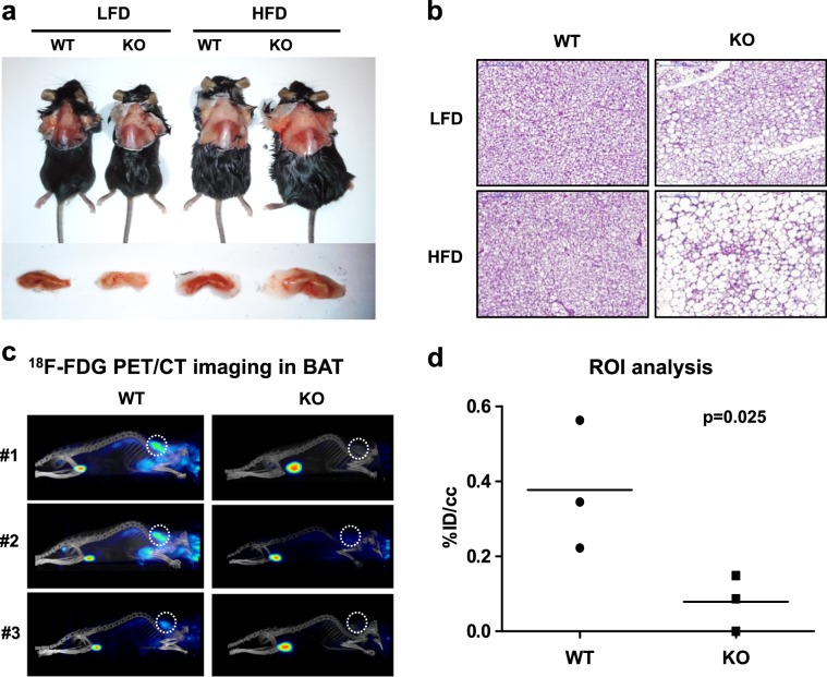
Fig. 2. IDH2 deficiency leads to BAT whitening.
a Representative images showing interscapular brown adipose tissue (iBAT) deposits (n = 6 per group). b Hematoxylin and eosin (H&E) staining of the iBAT section (5 μm) from the WT or IDH2KO mice exposed to a high-fat diet for 4 weeks. c PET/CT images showing the 18F-FDG uptake in the iBAT of the WT and IDH2KO groups as described in the “Materials and methods” (n = 5 per group). d Quantitative positron emission tomography–computed tomography (PET/CT) region of interest (ROI) analysis of the 18F-FDG uptake in the iBAT; 18F-FDG: 2-deoxy-2-[18F]-fluoro-d-glucose.

