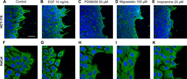
Fig. 5. Cell morphology of HCT-116 and HaCat cells upon inhibition of lamellipodia and filopodia formation.
Inset shows the immunofluorescence analysis of the fascin1 marker (green) under control conditions: a–f control conditions, b–g 10 ng/mL EGF (migration stimulator), c–h 50 µM PD98059 (MEK inhibitor), d–i 100 µM migrastatin, and e–k 20 µM imipramine. Images were captured using an LSM 510 META confocal fluorescence microscope with a ×63 oil objective. Scale bar 30 µm. Migrastatin and imipramine inhibit lamellipodia protrusion and fascin1 localization in a similar way to the migration MEK inhibitor PD98059 in both cell lines.

