Fig. 1.
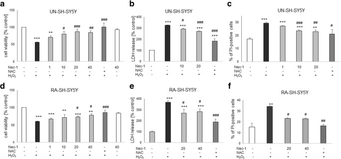
The effect of necrostatin-1 on H2O2-induced cell damage in UN- and RA-SH-SY5Y cells. UN- and RA-SH-SY5Y cells (a–c and d–f, respectively) were pre-treated for 30 min with necrostatin-1 (Nec-1; 1–40 μM) followed by 24 h of treatment with H2O2 (0.25 and 0.5 mM for UN- and RA-SH-SY5Y, respectively). As a positive control for the assays, we used antioxidant N-acetylcysteine (NAC, 1 mM) which was given concomitantly with the cell damaging factor. a, d Results of cell viability assessment in UN-(a) and RA-(d) SH-SY5Y cells measured by the MTT reduction assay. Data were normalized to vehicle-treated cells (control) and are presented as the mean ± SEM from 3 to 11 separate experiments with 5 repetitions each. (b, e) Results of cell toxicity assessment in UN-(b) and RA-(e) SH-SY5Y cells measured by the LDH release assay. Data were normalized to vehicle-treated cells (control) and are presented as the mean ± SEM from 4 to 11 separate experiments with 5 repetitions each. c, f Flow cytometry results of propidium iodide (PI)-stained UN- (c) and RA (f) SH-SY5Y cells after 24 h of cell treatment. Data are presented as the mean ± SEM of PI-positive cells from 3 to 5 independent experiments with 2 replicates. **P < 0.01 and ***P < 0.001 vs. vehicle-treated cells; #P < 0.05, ##P < 0.01, and ###P < 0.001 vs. H2O2-treated cells

