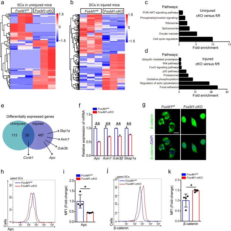
Fig. 3. Loss of FoxM1 results in activation of wnt/β-catenin signaling in SCs.
a, b Heatmaps showing the differential expressed protein-coding genes of SCs in uninjured mice (a) or injured mice (b) (n = 3). SCs were isolated from 2-month-old FoxM1-cKO mice that were uninjured or 48 h post-injury by flow cytometry. The sorted SCs were conducted by RNA-Sequencing analysis. c, d Enrichment of differentially expressed genes into pathways of SCs in uninjured mice (c) or injured mice (d). e Numbers of dysregulated protein-coding genes (data from RNA-Sequencing) in SCs. f SCs were sorted from FoxM1fl/fl and FoxM1-cKO mice (2 months of age) at 48 h post-injury. Total RNA of sorted SCs was extracted and the expression of the indicated genes was assayed by qPCR (n = 4). g SCs were isolated from FoxM1fl/fl and FoxM1-cKO mice (2 months of age) and cultured in vitro. Immunofluorescence staining of β-catenin in SCs (n = 3). Nuclei were stained with DAPI. Scale bars, 20 μm. h The mononuclear cells were isolated from the skeletal muscles of mice (2 months of age) at 48 h post-injury and were stained with SC markers and Apc fluorescent antibody. The intensity of Apc was analyzed by flow cytometry. i The mean fluorescence intensity (MFI) analysis of Apc in SCs (n = 4). j The mononuclear cells were isolated from the skeletal muscles of mice (2 months of age) at 48 h post-injury and were stained with SC markers and β-catenin fluorescent antibody. The intensity of β-catenin was analyzed by flow cytometry. k The MFI analysis of β-catenin in SCs (n = 4). Error bars represent the means ± SD. *p < 0.05, **p < 0.01; Student’s t test.

