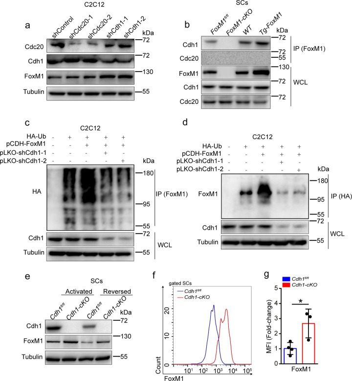
Fig. 6. Cdh1 prevents accumulation of FoxM1 protein by ubiquitylation.
a Cdc20 and Cdh1 were knocked down in proliferative C2C12 using independent shRNAs (pLKO construct). Levels of Cdc20, Cdh1, FoxM1, and Tubulin (loading control) were determined by western blot analysis (n = 3). b SCs were isolated from FoxM1 deletion or overexpression mice and were cultured in vitro. Cdh1 proteins were immunoprecipitated from the cell lysates and analyzed by western blot (n = 3). WCL whole-cell lysates. c, d HA-ubiquitin (HA-Ub, ubiquitin expression construct with HA tag) plasmid was transfected with the indicated plasmids (FoxM1 overexpression plasmid, pCDH-FoxM1; Cdh1 knockdown plasmid, pLKO-shCdh1-1, pLKO-shCdh1-2) in proliferative C2C12 cells; 8 h after 10 μM MG132 treatment, HA-Ubiquitin (c) or FoxM1 (d) proteins were immunoprecipitated from cell lysates and analyzed by western blot (n = 4). WCL whole-cell lysates. e SCs were isolated from Cdh1fl/fl and Cdh1fl/flPax7-Cre (called Cdh1-cKO here) and were cultured in growth medium (activated state) or serum-free medium (reversed state) in vitro. Whole-cell lysates were subjected to western blot analysis using the indicated antibodies (n = 3). f The mononuclear cells were isolated from skeletal muscles of mice (2 months of age) and were stained with SC markers and FoxM1 fluorescent antibody. The intensity of FoxM1 was analyzed by flow cytometry. g The MFI analysis of FoxM1 in SCs (n = 3–4). Error bars represent the means ± SD. *p < 0.05; Student’s t test.

