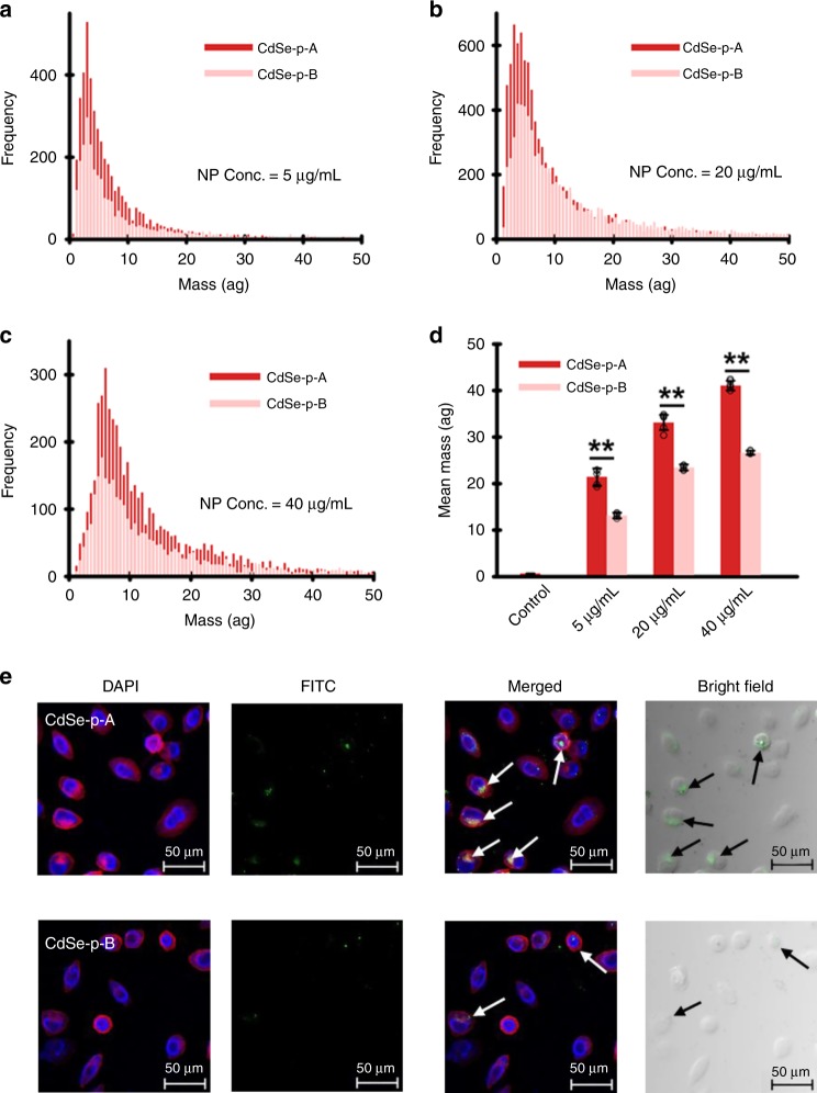
Fig. 3. Facet-dependent binding of transferrin and cellular uptake of CdSe nanoparticles.
a–d Single-cell–inductively coupled plasma–mass spectrometry (SC–ICP–MS) histogram of mass distribution (a–c) and mean mass (d) of CdSe nanoparticles (CdSe-p-A: dark red bar; CdSe-p-B: light red bar) taken up by HeLa cells, after cell incubation with transferrin–CdSe conjugates for 3 h. Results show higher cellular content of nano-Cd in HeLa cells exposed to “A” materials (i.e., CdSe with more (100) facet) that preferentially bound with transferrin. Data are presented as mean ± SD of five replicate samples (n = 5; p = 0.0007, 0.0004, <0.0001 for the group of 5, 20, 40 µg/mL CdSe nanoparticles by one-way ANOVA, respectively). e Confocal laser scanning microscopy images of HeLa cells after incubated with FITC-labeled transferrin–CdSe conjugates demonstrated greater cellular uptake of “A” materials. Microscopic analysis was independently repeated five times, and the results were similar. Statistical significance between groups: (**) p < 0.01. Source data are provided as a Source Data file.

