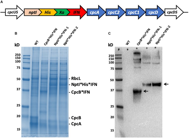
FIGURE 13.
(A) Map of the nptI∗IFN fusion construct in the cpc operon locus. Note the presence of the His-tag and the Xa protease cleavage site in-between the two genes in the fusion. (B) SDS-PAGE and Coomassie staining of the protein extracts from wild type (WT), the cpcB∗His∗IFN, and two independent lines of the nptI∗His∗IFN transformants. (C) Western blot analysis of a duplicate gel as the one shown in (B). Specific anti-IFN polyclonal antibodies were used in this analysis. Note the specific antibody cross reactions with protein bands migrating to ∼36 kD (CpcB∗His∗IFN) and ∼46 kD (NptI∗His∗IFN). Also note the antibody cross reactions with protein bands of higher molecular mass.

