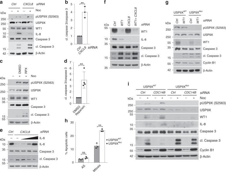
Fig. 4. CDK1/CDC14B-dependent phosphorylation of USP9X at serine 2563 promotes mitotic survival via WT1 and IL-8.
a Mitotic apoptosis in response to CXCL8 versus control knockdown detected by immunoblot in U2OS cells that were treated with the respective siRNA and arrested in mitosis using nocodazole for 8 h. Samples were collected, lysed, and analyzed by western blot with the indicated antibodies. b Quantification of relative amount of cleaved caspase 3 in n = 3 biologically independent experiments conducted as described in a. Ratio paired t-test was applied with **p = 0.0055. c Immunoblot analysis showing increased mitotic apoptosis in U2OS cells in response to treatment with nocodazole (8 h) and the CXCR1/2 inhibitor reparixin (RPX) or DMSO for 48 h. d Quantification of relative amount of cleaved caspase 3 in n = 4 biologically independent experiments conducted as described in c. Ratio paired t-test was applied with **p = 0.0031. e Immunoblot analysis confirming reversal of mitotic apoptosis following exogenous reconstitution of IL-8 in CXCL8-depleted cells. Experiment was performed as in a, with addition of exogenous IL-8 for the last 48 h. Cells were treated with nocodazole for 8 h. f Immunoblot analysis detecting induction of mitotic apoptosis in response to WT1, CXCL8, or WT1 and CXCL8 knockdown compared to control knockdown in U2OS cells that were arrested in mitosis using nocodazole (8 h). g Immunoblot analysis showing increased mitotic apoptosis by WT1 knockdown only in USP9XWT but not in USP9XMut U2OS cells. Analyzed cells were treated with control or WT1 siRNA and then arrested in mitosis using nocodazole. h Induction of mitotic apoptosis in USP9XMut U2OS cells that were kept asynchronous or treated with nocodazole for 32 h, stained with PI and measured by flow cytometry. PI-positive cells were quantified in each sample using FlowJo software. Mean is shown from n = 3 biologically independent experiments. Paired t-test was applied with **p(Mitosis) = 0.00278, p(AS) = 0.0745. i Immunoblot analysis revealing decreased mitotic apoptosis after CDC14B knockdown in USP9XWT but not in USP9XMut U2OS cells. Before lysis cells were transfected with control or CDC14B-directed siRNA, arrested in mitosis, and collected for analysis by western blot. Throughout this figure, mean and standard deviations as error bars are displayed.

