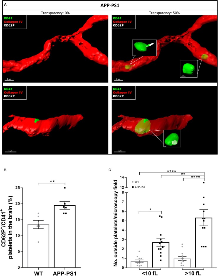
FIGURE 3.
Activation status of platelets located in the brain of APP-PS1 mice. Immunohistochemical analysis revealed that CD41+ platelets (green) expressing the activation marker CD62P (white) circulate within cerebral blood vessels (red) in APP-PS1 mice (A, upper row). Similarly, platelets in the process of fenestrating the vessel wall also expressed CD62P (A, lower row). Quantitative analysis showed that APP-PS1 mice have a significantly higher percentage of CD62P+/CD41+ platelets in the brain compared to WT animals (B). Moreover, in APP-PS1 mice, extravascular platelets exhibit a shift from single cells (<10 fL) to aggregates (>10 fL). Graph bars represent mean ± SEM (n = 6/group including WT: six females and APP-PS1: three females and three males). Statistical analysis was performed by unpaired Student’s t test (B) or two-way ANOVA with Tukey’s multiple comparison test (C). *p < 0.05; **p < 0.01; and ****p < 0.0001. Scale: 5 μm (A) and 1 μm (A—zoom in).

