FIGURE 5.
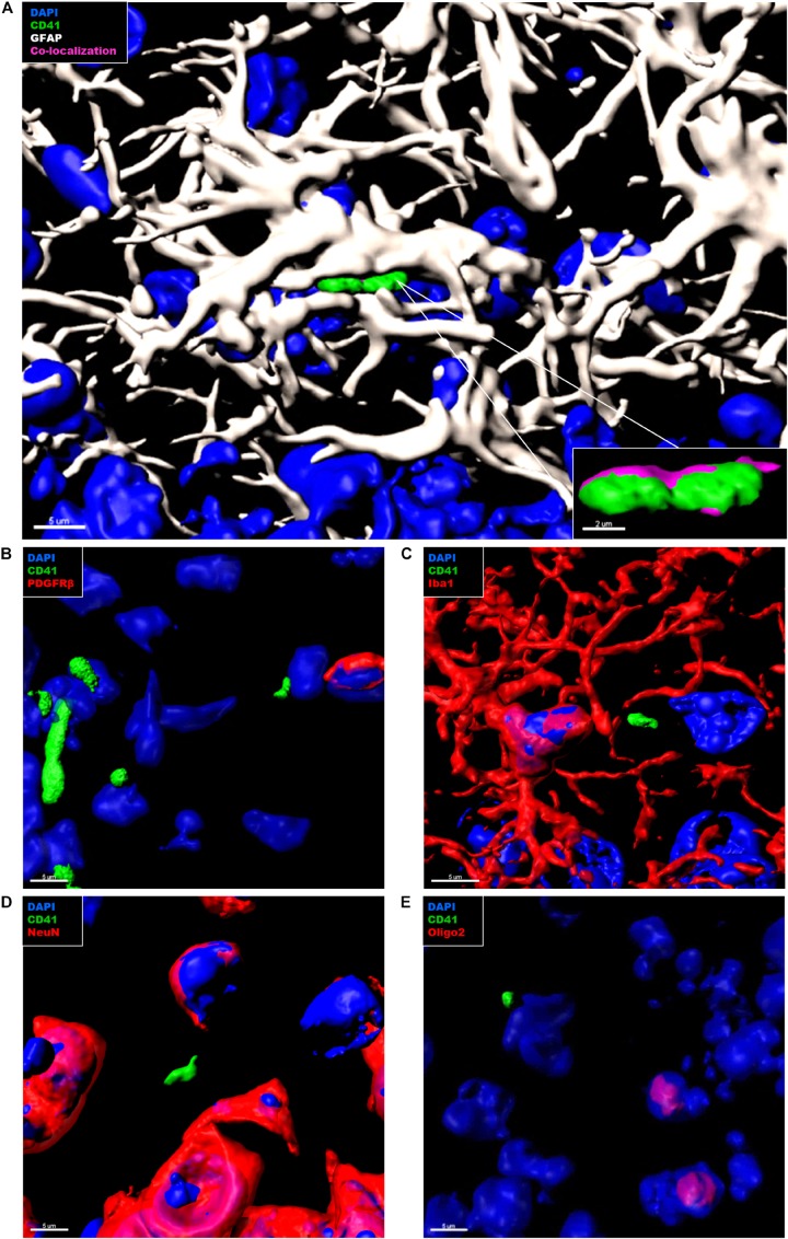
Immunohistochemical analysis of platelet interaction with neurovascular niche cellular components in the brain of APP-PS1 mice. Brain sections from APP-PS1 mice (n = 3, including two females and one male) were co-stained for platelets (CD41), (A–E, green), and one of the following cell types: astrocytes (GFAP) (A, white); pericytes (PDGFRβ) (B, red), microglia (Iba-1) (C, red), neurons (NeuN) (D, red), or oligodendrocytes (Oligo2) (E, red). DAPI (blue) was used as nucleus stain. Representative confocal images were selected for 3D surface projection and co-localization analysis. In APP-PS1 mice, platelets were found in tight association with astrocytes (A). Co-localization of CD41 and GFAP signals is depicted in magenta (A, zoom in). No further interactions were observed between CD41+ platelets and other immunolabeled cells (B). Scale: 5 μm (A,B), 2 μm (B—zoom in).

