Figure 3.

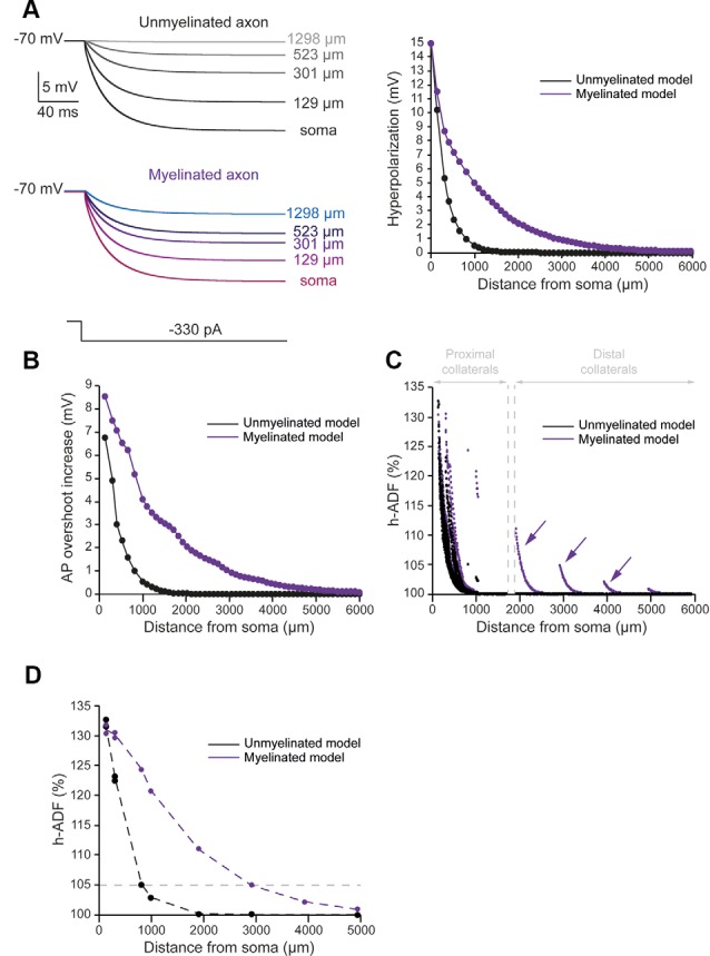
Axonal myelination increases h-ADF spatial extent. (A) Somatic subthreshold hyperpolarization propagates farther in the myelinated axon than in the unmyelinated axon. Left, voltage traces showing the propagation of the somatic hyperpolarization all along the axon (unmyelinated model: black to gray traces, myelinated model: purple to blue traces). Right, plot of the hyperpolarization in the function of the distance from the soma in the unmyelinated (black) and the myelinated (purple) model. (B) Plot of the hyperpolarization-induced AP overshoot increase in function of the distance from the soma in the unmyelinated (black) and the myelinated (purple) model. (C) Plot of the hyperpolarization-induced synaptic facilitation (h-ADF) in the function of the distance from the soma in the 1,982 presynaptic sites located in axon collaterals (black dots: unmyelinated model, red dots: myelinated model). Note that h-ADF is present in presynaptic sites located into distant collaterals only in the myelinated model (purple arrows). (D) Plot of h-ADF measured in the first presynaptic site of each collateral in the function of the distance from the soma (black: unmyelinated model, purple: myelinated model).
