Figure 4.

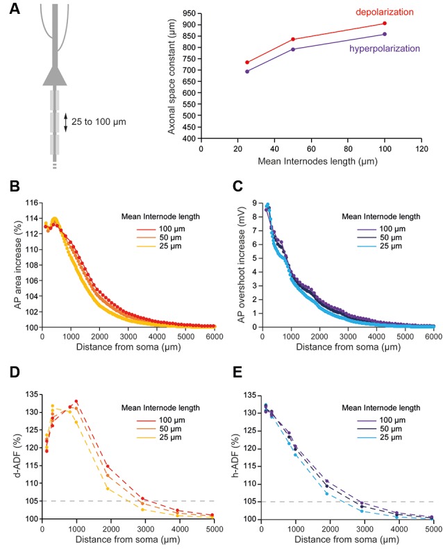
Internodal length mildly determines ADFs spatial extent. (A) The reduction of the internodal length leads to a decreased axonal length constant. Left, schematic representation of the model with varying average internodal lengths. Right, plot of the axonal length constant for a 10 s somatic depolarization (red) or a 200 ms somatic hyperpolarization (purple) in the function of the internodal length. (B) Plot of the depolarization-induced AP area increase in function of the distance from the soma for the different average internodal lengths. (C) Plot of the hyperpolarization-induced AP overshoot increase in function of the distance from the soma for the different average internodal lengths. (D) Plot of d-ADF measured in the first presynaptic site of each collateral for the different internodal lengths. (E) Plot of h-ADF measured in the first presynaptic site of each collateral for the different internodal lengths.
