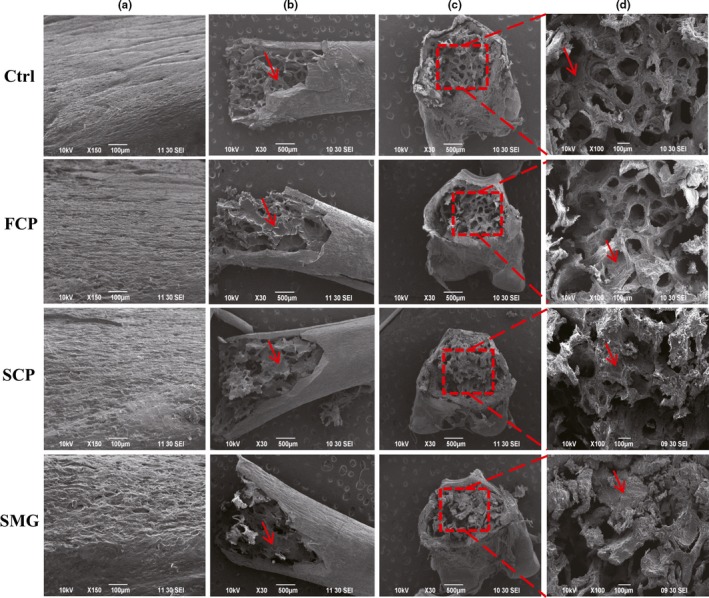
Figure 3.

Effects of FCP, SCP, and SMG on bone strengthening detected by scan electron microscope. (a): the surface of middle part of tibia shaft; (b): the cancellous substance in PTM; (c): the cross section of PTM; and (d): magnified image of corresponding red dashed boxes in c. The bone marrows are marked by red arrow
