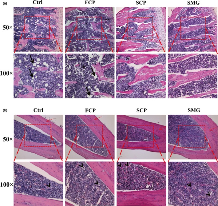
Figure 4.

Bone‐strengthening effects of FCP, SCP, and SMG in HE staining assay. (a): sections from PTM. (b): sections from middle tibia shaft. Pictures were taken at two kinds of magnification, 50 × and 100×. The 100 × pictures were obtained by magnifying the image in red dashed frames of corresponding 50 × pictures. Osteocytes are marked by red arrow. Adipocytes are marked by black arrow. Megakaryocytes are marked by black arrowhead
