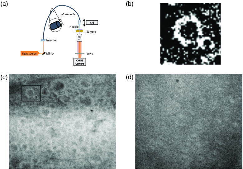
Fig. 4.
Possible transmission of light through myelin sheaths in thick longitudinal slices of spinal cord. (a) Optical arrangement for the imaging experiment. Light was collected on the underside of the slice by a objective and recorded by a CMOS cameras (DMK 23UP031, imaging source). (b) Magnified and contrasted image of bright myelin sheath and dark axon center from (c). (c) longitudinal slice illuminated using a 594-nm fiber laser source (MBL-FN, Changchun New Industries Optoelectronics Technology, China). Inset square shows location of zoomed image in (b). (d) 1-mm longitudinal slice illuminated with a fibered white lamp source (SLS201L, Thorlabs, New Jersey).

