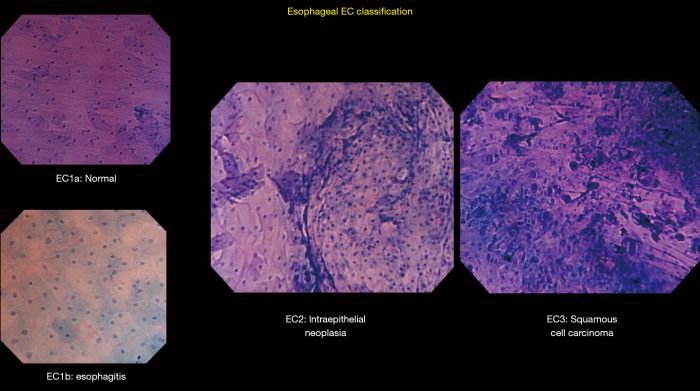
Figure 2.
Esophageal EC classification: representative pictures differentiating EC1a (normal), EC1b (esophagitis), EC2 (intraepithelial neoplasia), and EC3 (squamous cell carcinoma). EC1a shows regularly arranged large rhomboid-shaped cells. EC1b shows blunted edges and more rounded cells. EC2 shows an increase in cellular density but still with a recognizable cell structure. EC3 shows complete loss of cellular structure with a significant increase in cellular density.

