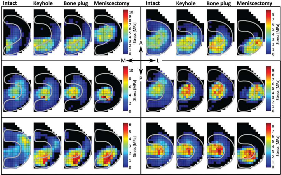
Figure 2.
Contact stress maps for each knee and each test condition at 45% of the gait cycle. Despite variability among knee contact patterns, a significant decrease in contact area and increase in peak contact stress in the cartilage-meniscal region of interest are shown for the meniscectomized condition. The decrease in contact area and increase in peak contact stress are also shown for the cartilage-cartilage region of interest for the meniscectomized condition as is the increase in peak contact stress for the bone plug condition. A, anterior; L, lateral; M, medial; P, posterior.

