
Fig. 2.
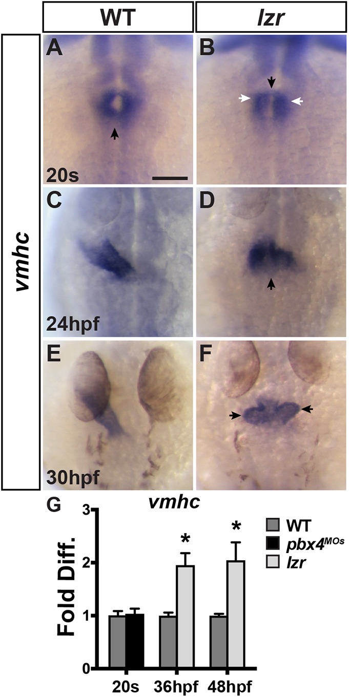
Cardiac fusion and elongation are abnormal in lzr mutants. (A-F) ISH for vmhc in WT and lzr mutant hearts at the 20 s stage, 24 hpf and 30 hpf. Views are dorsal with anterior up. Black arrows in A and B indicate the location of cardiac fusion when forming the cone. White arrows in B indicate anterior aggregates of CMs. Arrow in D indicates the larger ventricular pole. Arrows in F indicate already visible ventricular protrusions. At least 48 embryos per developmental stage were examined and genotyped. Scale bar: 100 µm. (G) RT-qPCR for vmhc expression in WT and Pbx4-depleted embryos at 20 s and in WT and lzr mutants at 36 hpf and 48 hpf. Error bars indicate s.e.m. *P<0.05.