Fig. 3.
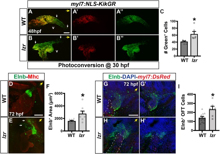
lzr mutants have an increase in later-differentiating ventricular CMs and SHF-derived smooth muscle. (A-B″) Confocal images of hearts from WT and lzr mutant myl7:NLS-KikGR embryos at 48 hpf following photoconversion at 30 hpf. White arrows indicate the area encompassing green-only cells within the OFT that was quantified. v, ventricle. (C) Number of green-only cells in WT (n=7) and lzr (n=6) myl7:NLS-KikGR hearts at 48 hpf following photoconversion. (D,E) IHC for Elnb (green) and Mhc (red) on hearts of WT and lzr embryos at 72 hpf. White arrows indicate extensions of Elnb in lzr mutant embryos. (F) Area (µm2) of Elnb expression in the OFT of WT (n=7) and lzr mutant (n=8) embryos. (G-H′) The OFT from hearts of WT and lzr mutant myl7:DsRed2-NLS embryos at 72 hpf. Elnb (green), DAPI (blue). Dashed line outlines the area containing CMs (red). G′ and H′ show the sections used to count Elnb-surrounded nuclei. Dots indicate representative nuclei. (I) Number of Elnb-surrounded cells in the OFT of WT (n=12) and lzr mutant (n=6) embryos. Yellow arrows indicate the direction of the arterial pole of the heart. Scale bars: 50 µm. Error bars indicate s.e.m. *P<0.05.

