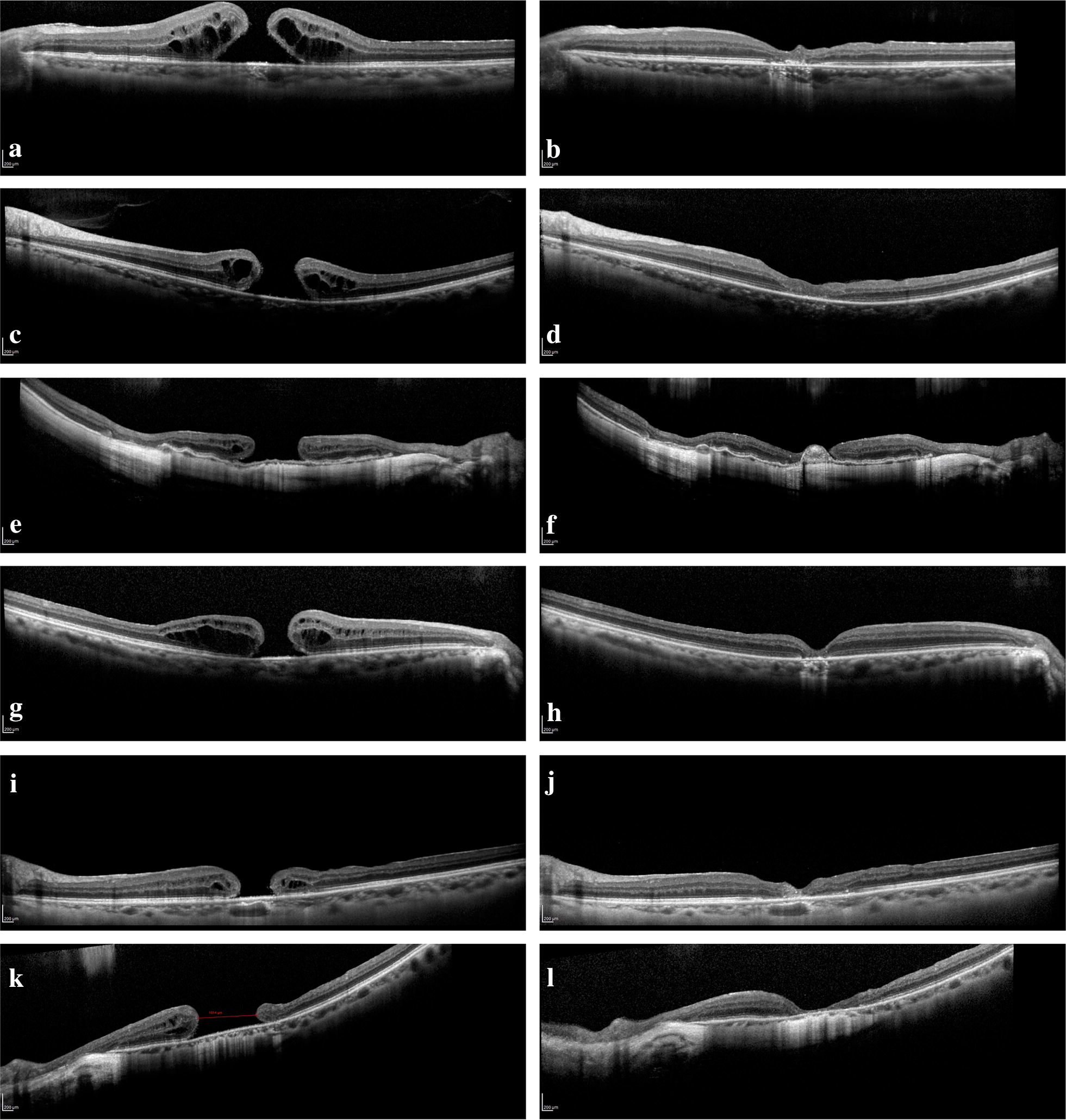
Fig. 1.

a–d Patient 1 and 2 with pre op OCT showing type 1 holes and post op OCT showing a closed macular hole. e, f Patient 3 with pre op OCT showing a type 3 hole and post op OCT showing ILM material but no neuroretinal tissue overlying the smaller hole (failed closure). g, h Patient 6 with pre op OCT showing a type 1 hole and post op OCT showing a closed macular hole. i, j Patient 7 with pre op OCT showing a chronic (1333 days) type 3 hole (617 μm) with closure seen on the post op OCT. k, l Patient 8 with pre op OCT showing a large (1014 μm), chronic (1481 days) type 3 hole with closure on the post op OCT showing a thin layer of overlying continuous neuroretinal layer 209 × 296 mm (300 × 300 DPI)
