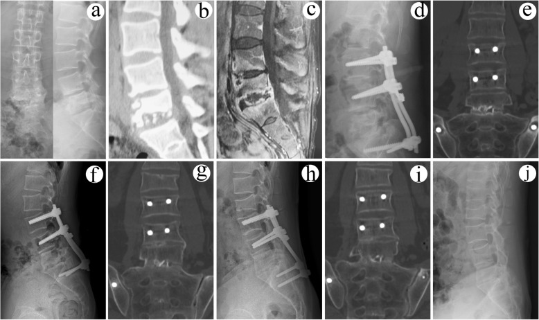
Fig. 4.
A 28-year-old male with L5/S1 lesions was managed by surgery with combined posterior-anterior approaches. The preoperative imaging data (a X-ray, b CT, and c MRI) showed severe vertebral destruction at L5/S1. The postoperative (d) radiography showed that internal fixation instrument was in good position. e CT and f X-ray indicated solid bony fusion at 12 months after surgery. At the 48 months after surgery, g CT and h radiography showed solid bony fusion without signs of fixation failure. At the final follow-up (96 months after surgery), i CT illustrated solid bony fusion and no obvious angle loss with good fixation position. j The radiograph showed the removal of internal fixation instrument and satisfied sagittal sequence

